Evaluating the impact of artificial intelligence-assisted image analysis on the diagnostic accuracy in detecting fractures on plain X-rays (FRACT-AI protocol)
Objective:
Missed fractures are the most frequent diagnostic error attributed to clinicians in UK emergency departments and a significant cause of patient morbidity. Recently, advances in computer vision have led to artificial intelligence (AI)-enhanced model developments, which can support clinicians in the detection of fractures. Previous research has shown these models to have promising effects on diagnostic performance, but their impact on the diagnostic accuracy of clinicians in the National Health Service (NHS) setting has not yet been fully evaluated.
Methods:
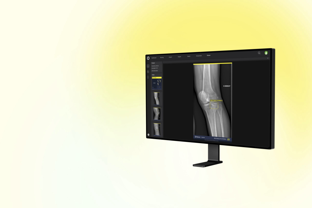
A dataset of 500 plain radiographs derived from Oxford University Hospitals (OUH) NHS Foundation Trust will be collated to include all bones except the skull, facial bones and cervical spine. The dataset will be split evenly between radiographs showing one or more fractures and those without. The reference ground truth for each image will be established through independent review by two senior musculoskeletal radiologists. A third senior radiologist will resolve disagreements between two primary radiologists. The dataset will be analysed by a commercially available AI tool, BoneView (Gleamer, Paris, France), and its accuracy for detecting fractures will be determined with reference to the ground truth diagnosis. We will undertake a multiple case multiple reader study in which clinicians interpret all images without AI support, then repeat the process with access to AI algorithm output following a 4-week washout. 18 clinicians will be recruited as readers from four hospitals in England, from six distinct clinical groups, each with three levels of seniority (early-stage, mid-stage and later-stage career). Changes in the accuracy, confidence and speed of reporting will be compared with and without AI support. Readers will use a secure web-based DICOM (Digital Imaging and Communications in Medicine) viewer (www.raiqc.com), allowing radiograph viewing and abnormality identification. Pooled analyses will be reported for overall reader performance as well as for subgroups including clinical role, level of seniority, pathological finding and difficulty of image.
This study is registered with ISRCTN (ISRCTN19562541) and ClinicalTrials.gov (NCT06130397).


